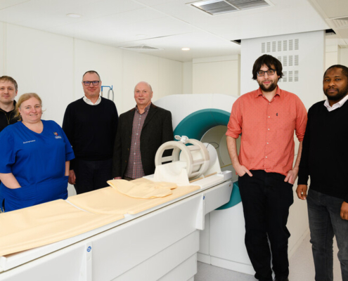
We are supporting the University of Aberdeen with a study to trial the use of next-generation imaging technology to detect declining brain health earlier.
The Field‑Cycling Imaging (FCI) scanner has already been shown to be highly effective in producing previously unattainable imaging following stroke, identifying cancer spread and brain tumours. This new study could now transform how clinicians detect the earliest signs of declining brain health.
The team at the University will initially focus on cerebral small vessel disease, a common age‑related brain condition that increases the risk of stroke and dementia. Although most people develop some degree of small vessel disease over time, identifying who is at risk of severe decline remains a major clinical challenge. Unlike conventional MRI, FCI can switch between low and ultra‑low magnetic fields, revealing disease effects that were previously impossible to see.
Funded by the Scottish Government Chief Scientist Office and NHS Grampian Charity, the team will investigate how effectively FCI can detect changes in brain health as we age with a view to identifying those who could most benefit from early intervention treatment which could slow this decline.
We are supporting the project through raising awareness of the study with our IONA cohort in Aberdeen.

Innovative technology
Field cycling imaging (FCI) is a new and specialist type of low-field MRI scan pioneered in Aberdeen. The FCI scanner follows in the footsteps of the full body MRI scanner, invented at the University around 50 years ago which has gone on to save millions of lives around the world. The FCI derives from MRI but can work at low and ultra-low magnetic fields which means it is capable of seeing how organs are affected by diseases in ways that were previously not possible.
The project will bring together a wealth of scanning expertise from the University using a variety of brain‑imaging methods. Some volunteers will also have a low‑field MRI scan at the Centre for Adaptable Imaging Technologies, using techniques developed by Research Postgraduate student Gabriel Zihlmann and Dr Mathieu Sarracanie. This part of the study aims to see whether what is learned from FCI can also be picked up by low‑field MRI, helping researchers explore ways to create more affordable and portable MRI tools for checking brain health as people get older.
Comments from the researchers and supporters
Dr Gordon Waiter, Reader and director of University of Aberdeen Biomedical Imaging Centre, who is co-leading this research study:
“With people living longer it is becoming more pressing that we understand the earliest stages of declining brain health. In Scotland we are excellently placed to take on this challenge. With support from the Chief Scientists Office of the Scottish Government, NHS Grampian Charity and Scottish Brain Sciences we will use a unique imaging technology developed here at the University of Aberdeen to investigate brain health and help to inform future treatments. However, this project could not go ahead without the people of the North East of Scotland. Volunteers are a crucial part of our work and our collaboration with Scottish Brain Sciences means we can involve the local population in research that will benefit all of Scotland.”
“This new arm of research at the University of Aberdeen is underpinned by a talented team of early career researchers working across imaging physics and computer science.
“Our results from the previous PUFFINS2 project which used the previous generation of FCI scanner have already shown that FCI can detect the presence of moderate and severe cerebral small vessel disease. The objective of this new study is to use the improved performance of the next generation FCI scanner to determine whether and to what extent FCI can detect earlier disease, and the progression of disease over time.”
Prof Craig Ritchie, CEO and Founder of Scottish Brain Sciences:
“We’ve been delighted to be able to support this important research from our home at BioHub through signposting participants in our IONA Cohort to the work. This is a fantastic example of the ’triple helix’ in action where the Life Sciences sector, University and NHS all work in concert to achieve impactful outcomes. I’d hope this is the first of many such collaborations here in Aberdeen.”
Prof Mary Joan MacLeod, Chair at The Institute of Medical Sciences at the University of Aberdeen, who is co-leading this research study:
“It’s exciting that Aberdeen is once again at the forefront of advances in imaging. We hope this study will show that this technology will make it easier to screen and identify the population who are at risk of developing dementia, so that treatment can be given early.”
Lisa Duthie, NHS Grampian Charity Lead:
“This innovative research has the potential to transform how changes in brain health are detected, which could have significant implications in the diagnosis and treatment of conditions like stroke and dementia in future.
“We’re proud to support the research team to carry out this work, and we are very grateful to all the volunteers whose participation is absolutely vital in making research like this possible.
“This study has real potential to make a lasting difference to people here in Grampian, and further afield, who are at risk of cerebral small vessel disease and the conditions it contributes to.”
More news from Scottish Brain Sciences
 https://brainsciences.scot/wp-content/uploads/2026/02/FCi_brain_study_group.jpg
533
800
Denise Fraser
https://brainsciences.scot/wp-content/uploads/2025/11/SBS-web-logo5-1.png
Denise Fraser2026-02-16 15:56:592026-02-16 16:07:17Championing new technology to detect declining brain health earlier
https://brainsciences.scot/wp-content/uploads/2026/02/FCi_brain_study_group.jpg
533
800
Denise Fraser
https://brainsciences.scot/wp-content/uploads/2025/11/SBS-web-logo5-1.png
Denise Fraser2026-02-16 15:56:592026-02-16 16:07:17Championing new technology to detect declining brain health earlier https://brainsciences.scot/wp-content/uploads/2026/01/Group-shot-scaled.jpg
1707
2560
Denise Fraser
https://brainsciences.scot/wp-content/uploads/2025/11/SBS-web-logo5-1.png
Denise Fraser2026-01-29 10:32:272026-01-29 10:32:46Aberdeen FC partnership shines a spotlight on brain research in the north east
https://brainsciences.scot/wp-content/uploads/2026/01/Group-shot-scaled.jpg
1707
2560
Denise Fraser
https://brainsciences.scot/wp-content/uploads/2025/11/SBS-web-logo5-1.png
Denise Fraser2026-01-29 10:32:272026-01-29 10:32:46Aberdeen FC partnership shines a spotlight on brain research in the north east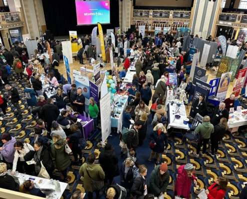 https://brainsciences.scot/wp-content/uploads/2025/12/Festival-2025-photo.jpg
1200
1600
Denise Fraser
https://brainsciences.scot/wp-content/uploads/2025/11/SBS-web-logo5-1.png
Denise Fraser2025-12-04 10:26:522025-12-04 11:45:35Talking brain health at Aberdeen Wellbeing Festival
https://brainsciences.scot/wp-content/uploads/2025/12/Festival-2025-photo.jpg
1200
1600
Denise Fraser
https://brainsciences.scot/wp-content/uploads/2025/11/SBS-web-logo5-1.png
Denise Fraser2025-12-04 10:26:522025-12-04 11:45:35Talking brain health at Aberdeen Wellbeing Festival